Journal of Advanced Veterinary Research

Volume 9, Issue 3, 2019, Pages: 117-122


(ISSN: 2090-6277/2090-6269/ © 2011-2019 JAVR)

www.advetresearch.com


Ultrasonographic, Morphometric and Histological Study of Testicular Parameters in Egyptian Water Buffalo Bulls (Bubalus Bubalis)


Tamer M. Genedy1*, Seham S. Hadad2, Emad M. Abd El-Razek1


1Department of Theriogenology, Faculty of Veterinary Medicine, University of Sadat City, Egypt.

2Department of Anatomy and Embryology, Faculty of Veterinary Medicine, University of Sadat City, Egypt.


Received: 13 June 2019; Accepted: 3 July 2019


*Corresponding author: Tamer M. Genedy (tamer.genedy@vet.usc.edu.eg)


Abstract


This study was carried out to identify testicular biometric data of native Egyptian buffalo-bulls through the determination of testicular parameters by using ultrasound examination of the testis and scrotum and determination of scrotal circumference of the life animal and morphometric data of the testis after slaughter of the bulls and their correlations with body weight at the different ages that will provide data base for the reproductive anatomy of the local Egyptian buffalo bull. The animals were divided into three groups (G1, G2 and G3) according to the age and the weight of the animals. All animals were examined with ultrasonography to determine the different parameters of the testis, epididymis and pampiniform plexus. After slaughter of the animals, the left and right testes were taken for morphometric and histological analysis. The results revealed significant (P˂0.05) variations between body weight and testicular parameters that included (Testicular Weight, Testicular Width, Testicular Circumference, Testicular Thickness and Testicular Length) as there was an increase in all parameters with the advancement in age and body weight. Histological analysis of G1 declared that only sertoli cells rested on basal lamina of seminiferous tubules and Gonocyt cells are predominant but there was no spermatogenic cells, no spermatocyte and no spermatid in the lumen of seminiferous tubules. In G2 group, there was replacement of Gonocyt cells with differentiated spermatogenic cell and spermatocyte in the lumen of seminiferous tubule but there was no spermatid. In G3 group, there were different stages of developmental spermatogenic cell clusters within the epithelium of seminiferous tubules (sertoli cells spermatogenia cells, different cells type of spermatocyte, and  round  spermatid as well as several threads like sperms were in the lumen of seminiferous tubules) that  indicate the early maturity of Egyptian  buffalo bull noticed at this age Statistical analysis revealed significant (P˂0.05) changes in the testicular parameters with the advancement in the age and weight of the animals concluding that ultrasonographic imaging of the testis and epididymis of buffalo-bulls was given appreciable benefits in studying the developmental changes of the testes and epididymis of buffalo-bulls, so that ultrasound examination of testicular parameters is a good tool for prediction of the future fertility of buffalo-bulls.

Keywords: Buffalo-bulls, Histological and morphometric, Testes, Ultrasound


Introduction


Buffaloes are an economically important source for meat and milk production, as well as work output in difficult condition better than cattle (Nascimento and Carvalho, 1993), and it characterized by their high fertility, longevity, feed conversion efficiency and productivity in comparison to cattle (Bernardes, 2007). Recently, there is an interest for reproduction and management in buffalo species as their high adaptive ability to tropical and subtropical climatic conditions and their capacity to survive in areas unsuitable for cattle and other domestic animals, (Patrícia et al., 2013).

Testicular biometry include testicular length (TL), testicular width (TWD), testicular thickness (TT), testicular weight (TW) and testicular circumference (TC) and scrotal circumference (SC) are very important elements for monitoring the testis normality and judging potential sperm production (Paula and Navarro, 2001), as there is high correlation and strong relationship between SC and testicular parameter, with age and body weight in Murrah Buffalo Bulls as well as reproductive capacity (libido) particularly sperm production (Patrícia et al., 2013). The biometric data related to testicular parameters and SC help in breeding selection and assist in reproduction to characterize puberty and sexual maturity and enable inferences about spermatogenesis (Patrícia, et al., 2013).

Ultrasonographic examination of reproductive system is an effective diagnostic clinical technique for differentiating potential bulls as it represented as complementing clinical examination (Gnemmi and Lefebvre 2009). Kahn (1994) added that ultrasonographic examination of reproductive system is useful in predicting many lesions and abnormalities of the reproductive tract. The main functions of ultrasound are evaluating anatomical structures and determining the echogenicity of testicular parenchyma (TP) and mediastinum (Chandolia et al., 1997; Clark, et al., 2003). Ultrasound also is useful in monitoring progressive developmental changes that occur in testis at different stages of maturation (Ahmad and Noakes, 1995).

This study was aimed to identify testicular biometric data for native Egyptian buffalo-bulls by determination of testicular parameters by using ultrasound examination of the testis and scrotum, measuring of scrotal circumference of the life animal and morphometric data of the testis after slaughter of the bulls and their correlations with body weight at the different ages that will provide data base for the reproductive anatomy of the local Egyptian buffalo bull.


Materials and methods


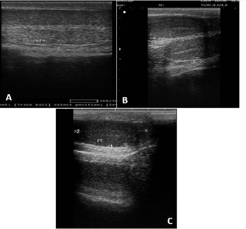
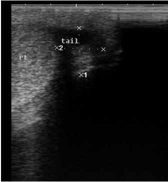
This study was conducted in the Department of Theriogenology in collaboration with Department of Anatomy and Embryology, Faculty of Veterinary Medicine, Sadat City University, Egypt.


Experimental location

The animals were reared in EL-Waddy farm on Cairo-Alexandria dessert road near to Sadat City. The city's climate is tropical with mild rainy winters and hot summers. The buffalo-bulls were fed barseem during winter season and supplemented with corn silage during the dry periods, water and mineral salt were added ad libitum. The buffalo-bulls used in this study were of different ages at the time of ultrasonographic examination. The buffalo-bull’s reproductive tracts (two tests with two epididymis and scrotum) were immediately collected after slaughtering from abattoir at Kom Hamada city and brought to the laboratory in ice for morphometric study and then specimens from the testicular parenchyma were preserved in 10% neutral buffer formalin and were processed on the same day.


Animals

          Fifteen Egyptian buffalo-bulls were divided into three groups: G1 (n =5) of 4-8 months’ age and 150-200 kg weight, G2 (n =5) of 8-12 months old and 200-250 kg weight and G3 (n = 5) of 12-16 months’ age and 300-350 kg weight. All animal-management procedures were carried out following the regulations of Institutional Animal Care Committee of Sadat City University, Egypt and in accordance with Egyptian ethical code for studies on experimental animals and Use Committee (IACUC) and with the prior approval for using the animals.


Ultrasonographic Examination

The animals were physically restrained in stanchion and they were weighted using a mechanical balance. Clinical examination of the animals was performed with a special attention to the general health of each buffalo bulls. The scrotum was observed for the presence of any lesions and the testes were palpated. Scrotal Circumference (SC) was evaluated using metric tape placed at the greatest diameter and measured as reported by (Osinowo et al., 1981). Free movement of the testis into the scrotum and lack of other tissue mass were confirmed. Ultrasonographic imaging of the testes was carried out where the animal was in standing position after clipping the hairs on the scrotum by using portable Ultrasonic Diagnostic System (Sonoscape Co. Ltd., China) provided with linear transducer (3.5-8 MHz). After securing the animal, gel was applied directly on the scrotum and then imaging was done. Longitudinal and transverse planes were imaged for scanning of testes. For imaging the longitudinal plane, mediastinum streak of each testis was taken as the landmark, while the dot mediastinum was taken as landmark for imaging the transverse planes. Images were frozen on the monitor of the ultrasound scanner and diameters of the testes were taken where the length of testes was measured in longitudinal plane and the width was measured perpendicular to the longitudinal plane (Jeyakumar et al., 2012). The width and the length of the pampiniform plexuses and the tail of epididymis were measured also.


Experimental Design


Testicular parameters (morphometric)

Weighting of the samples was done using a highly sensitive balance in the laboratory. After collection, the epididymis was separated from the testis. The left and right testes were measured separately and their weights were recorded. The testicular weight (TW), testicular length (TL), testicular width (TWD), testicular thickness (TT) and testicular circumference (TC) were determined for each testis. The length, width and thickness were measured using calipers with millimeter divisions. The TC and scrotal circumference (SC) were measured using a metric tape.


Histological examination

The middle portions of the testicular parenchyma obtained from 9 Egyptian buffalo bulls of different age and weight then, placed 10% formalin (for tissue fixation). Then dehydration in increasing concentrations of alcohol, followed by infiltration with xylenes, the tissue samples were embedded in paraffin. A microtome (Leica RM2145, Leica, Berlin, Germany) was used for sectioning the tissue into 5-mm-thick slices and these sections were stained with hematoxylin and eosin (HE) stains. The samples were analyzed under a light microscope (Leica DM 2500, Leica).


Statistical analysis

Descriptive analyses of the mean and standard error of means for each testicular biometric parameter were performed with the Graph Pad Prism4 software (Graph Pad Software, La Jolla, CA, USA). ANOVA was used for statistical analysis.


Results


Testicular parameters (morphometric)

There was a significant (P˂0.05) relation between body weight and testicular parameters that include (TW, TWD, TC, TT and TL) as there were increases in all parameters with the advancement in age and body weight (Table 1). The SC and body weight differed significantly between the three groups and there was an increase in the SC with increasing body weight (Table 2).


Table 1. Relation between testicular parameters and body weight, mean± SEM

Group

Body weight Kg

Right testis

Left testis

T W

(g)

T L

(cm)

T WD

(cm)

T T

(cm)

T C

(cm)

T W

(g)

T L

(cm)

T W

(cm)

T T

(cm)

T C

(cm)

1

150.00±5.77

18±0.57a

2.46±0.08a

1.16±.03a

1±0.05a

3.9±0.05a

18.67±0.66 a

2.46±0.06a

1.16±0.03 a

0.96±0.03 a

3.93±0.03 a

2

243.33±8.82

26±0.57b

3.13±0.08b

1.73±0.08b

1.5±0.05b

5.06±0.06b

25.67±0.88 b

3.1±1b

1.83±0.06 b

1.63±0.08 b

5.03±0.08 b

3

356.66±3.33

33.3±0.88c

6.1±0.06c

3.16±0.08c

2.9±0.08c

9.2±0.14c

34.33±0.33 c

6.1±0.15c

3.2±0.11 c

3.06±0.06 c

9.3±0.06 c

Values with different letters within each column were differed significantly (P<0.05)

TW = testicular weight, TL = testicular length, TWD = testicular width, TT = testicular thickness, TC = testicular circumference


Table 2. Ultrasonographic parameters of the testis, epididymis and pampiniform plexus

Groups

Body weight

(Kg)

SC

(cm)

Pamp. length

(mm)

Pamp. Breadth

(mm)

Right Testis

(mm)

left Testis

(mm)

Tail epid.

(mm)

Right

Left

Right

Left

L

B

L

B

Right

Left

1

150.00±5.77

15.33±1.2a

10.16 ±1.44a

10.33±0.33a

15±1.27a

17.9±0.3a

45.23±1.16a

16.36±0.17a

43.53±1.75a

19.03±1.23a

18.37±0.32a

18.1±0.15a

2

243.33±8.82

23±0.57b

19.2±2.35b

16.6±2.27b

23.66±1.94b

21.5±1.43b

55.40±0.73b

23.06±3.21b

55.8± 0.66b

31.6± 5.51b

20.6±1.23a

18.83±0.17a

3

356.66±3.33

26.66±0.66c

23.63±0.92c

30.73±1.32c

-

-

60.73±0.43c

44.36±2.72c

61.8±1.05c

38.16±3.3c

22.13±0.78a

22±1.15a

Values with different letters within each column were differed significantly (P<0.05)

Pamp. = Pampiniform plexus, L = Length, B = Breadth, Tail epid. = Tail of epididymis


Histological findings

Normal testicular parenchyma of buffalo bull was consisted of solid testicular cell cords in between them there were interstitial Leydig cells, vessels and nerves. There were numerous seminiferous tubules each one was surrounded by basement membrane and contains many cell clusters as sertoli cells, which were tall columnar cells resting on the basement membrane in mature Egyptian buffalo-bulls and other gonocyt cells that differentiated into spermatogenic cells and different type of spermatocyte in the lumen of seminiferous tubules and round spermatids as well as luminal spermatozoa.

The testis of immature buffalo bulls with an average body weight 150.00±5.77 kg showed different stages of cell development within the epithelium of seminiferous tubules, only appeared sertoli cells of round and circular shape that rest on basal lamina of seminiferous tubules and gonocyt cells were predominant but there was no spermatogenic cells, and no spermatocyte as well as there was no spermatid in the lumen of seminiferous tubules (Fig. 1A).

There was a marked variation in seminiferous tubules especially in their lumen of testis, Gonocyt cells were differentiated into spermatogenic cell and spermatocyte in the lumen of seminiferous tubule in buffalo bull with an average body weight 243.33±8.82 kg, but there is no any spermatid and no luminal sperms. (Fig. 1B).

While the testis of buffalo bulls with an average body weight 356.66±3.33 kg showed different stage of developmental spermatogenic cell clusters within the epithelium of seminiferous tubules (sertoli cells of tall shape cells, spermatogenia cells and different cell types of spermatocyte as well as there are round spermatids and luminal threads like sperms in the lumen of seminiferous tubules) so it is considered as be early mature animal at that age (Fig. 1C).


Fig. 1. Light photomicrograph of testis in Egyptian buffalo-bull with different body weights, blue arrows (nursing cell that called sertoli cells (ST) that resting on basement membrane of seminiferous tubules), L.C= Leydig or interstitial cells, SC = Spermatocyte with different of activity, SP = Spermatid in the lumen of the seminiferous tubules, G= Gonocyt.


Findings of the Ultrasonographic Scanning

Ultrasonographic imaging of the Egyptian buffalo-bull's testes revealed a homogenous hypoechoic parenchyma with centrally located hyperechoic mediastinum and surrounded by a distinct hyperechoic tunic (Fig. 2). There were significant (P<0.05) differences between the different ultrasonographic parameters of the testes of the three groups, as there was an increase in all parameters with the increase in age and body weight (Table 2).


Fig. 2. A) Cross section in testes width about 44.2mm the mediastinum appear and body of epididymis appear and hyperechoic tunics. B) Both testes in the same image. C) Right testes measures about 16.7 mm in breadth and 42.9 mm in length.


The tail of the epididymis was recorded from oblique plane near the distal pole of the testis. The tail is heterogeneous and less echogenic than testis (Fig. 3) although there was increase in measurements of the tail of the epididymis with increasing in age but no significant differences in epididymal tail diameter were recorded (Table 2).


Pampiniform plexus was appeared on the upper pole of testis where non-echogenic area containing scattered hyperechogenic spots (Fig. 4) and differed significantly between groups (P<0.05), its measurements increase significantly by advancement of age and weight (Table 2).


Fig. 3. Showing tail of epididymis of right testes about 19.1×18.2 mm


Fig. 4. Pampiniform plexuses of left side measures about 31.3mm in diameter


Discussion


Selection of buffalo bull for breeding required evaluation of its breed, body confirmations, libido and mating ability. Moreover, external and internal genitalia are examined and its semen quality must be considered. Biometric analyses are essential measurements in the andrological evaluation of a breeding animal. Okwun et al. (1996) reported that males with larger testis tend to produce more sperm. Testicular measurements and the changes that occur during growth of the testes from birth to maturity have been well documented for goats (Bitto and Egbunike, 2006), rams (Setchell, 1978) and bulls (Dyce et al., 2002). Both the scrotal circumference and body weight should be used together as a routine and reliable parameter to predict the reproductive capacity in buffalo (Carter et al., 1980). And (Ohashi et al., 2001) added that, the SC is one of the most commonly employed because it is easy to measure and highly correlated with body weight

There were many studies on the scrotal circumference of buffalo breed in the world. In the present study, the mean scrotal circumferences of Egyptian buffalo-bulls were 15.33±1.2 cm, 23±0.57 cm and 26.66±0.66 cm for G1, G2 and G3 respectively, which were appeared more than that of the pubertal swamp buffaloes (17-20 cm) which was reported by McCool and Entwistle (1989), that might be attributed to the breed difference, food availability and quality. The present study revealed a significant increase in SC with advancement of age and weight of the animal, which was similar to that reported in Murrah buffalo (Pant et al., 2003; Patrícia et al., 2013) and swamp buffalo, (Viana et al., 2008; Ohashi et al. (2001). The latter concluded that among the available biometrics for testes, SC is one of the most commonly employed because it is easy to measure and highly correlated with body weight.

Ultrasonography is one of the most widely used diagnostic tools in modern medicine used to visualize many internal organs (size, structure and any pathological lesions). Ultrasound permits a non-invasive evaluation of internal structure of scrotum and testis with its fibrous tunics, as well as parenchyma and mediastinum and follow their changes. In the current study, the testicular parenchyma showed changes in echogenicity of its ultrasonic image during development, this might be attributed to cellular proliferation and fluid production. At the same time, there was an increase in testicular echo-density as bulls achieved puberty which was agreed with that recorded by Chandolia et al. (1997).

In this study, the ultrasound findings of the testicular length and diameter as well as histological investigation were indicated a significant change with the advancement in the age and weight of the animal. , that is very clear in the difference between G.2 of 250 k.g B.w & G.3 of 350 K.g B.W., there were different type of spermatocyte activity or different stage of cellular association in the lumen of seminiferous tube, that is similar to that observed by (Curtis and Amann, 1981) in Holstein bulls .n the other hand, there was no significant change in the epididymal diameter this might be attributed to the early maturity of the animal without sexual use. 

In the current study, the tail of the epididymis was a more heterogeneous structure than the testis. As a whole, its structure was hypo-echogenic relative to that of the testis. These findings were comparable to those recorded by Cartee et al. (1986) and Pugh et al. (1990) for the boar and dog, respectively. Similar observations were recorded for bulls (Cartee et al., 1989), buck and rams (Ahmad et al., 1991; El-Baz and Abdel-Razek, 2019a, b). It seems that the testis was relatively more homogenous, whereas the tubules in the epididymis were larger and thus generate more specular echoes (Leung et al., 1984), which might have been responsible for its heterogeneous appearance. The head of the epididymis was less echogenic than the testicular parenchyma and homogeneous in echo-texture. Numerous hypoechoic tubular structures represented pampiniform plexus. The presence of significant relation in all parameters in the present study suggested that TL, TWD, TC and TT were useful parameters for the selection of the breeding animals.

The results of the present study were matched with that observed by Ahmad et al. (2010) and declared the establishment of spermatogenesis as reflected by the appearance of significant number of spermatids. In this study, buffalo bulls with 356 kg B.W (G.3), and of age 9-12 month, that was similar to that observed by Ohashi et al. (2001) showed the onset of the spermatogenic process in buffalo at 9 months. Thus, the establishment of spermatogenesis was progressive by the age and increasing in the body weight. On the other hand, at the same body weight of Egyptian buffalo bull (356 k.g), the obtained histological investigations of the spermatogenic process explained that these animals were early sexually mature due to presence of several threads like sperms in the lumen of seminiferous tubules. The spermatozoa was reported in the tubular lumen of swamp buffalo at 16 months (Bongso et al., 1984); In crossbred buffalo (Mediterranean × Jaffarabadi), at 13 months (Melo and Vale, 1992) and in Murrah Buffalo at 24 months (Patrícia et al., 2013). Under the husbandry conditions described here, both body and testicular development of buffalo-bulls occurred more slowly than the typical growth rates for Bos taurus (Coulter, 1991).


Conclusion


Biometric scrotal circumference and testicular parameters of Egyptian buffalo-bulls were useful indicators for the selection of bulls for breeding. Ultrasonographic imaging of the testis and epididymis of buffalo-bulls was given appreciable benefits in studying the developmental changes of the testes and epididymis with the advancement of the age and weight of buffalo-bulls so that, ultrasound examination of testicular parameters is a good tool for prediction of the future fertility of buffalo-bulls.


Conflict of Interests


The authors declare that there is no conflict of interest regarding the publication of this article.


References


Ahmad, N., Noakes, D.E., 1995. Ultrasonic imaging in determining the presence of testicular degeneration in two male goats. Br. Vet. J. 151, 101–109.

Ahmad, N., Noakes, D.E., Subandrio, A.L., 1991. B-mode, real time ultrasonographic imaging of the testis and the epididymis of sheep and goats. Vet. Rec. 128, 491-496.

Ahmad, N., Umair, S., Shahab, M., Arslan, M., 2010. Testicular development and establishment of spermatogenesis in Nili-Ravi buffalo bulls. Theriogenology 73, 20–25.

Bernardes, O., 2007. Buffaloes breeding in Brasil: position and economic relevancy. Rev. Bras. Reprod. Anim. 31, 293–298.

Bitto, I. I., Egbunike, G. N., 2006. Seasonal variations in the morphometric characteristics of the pubertal West African Dwarf Bucks in its Native Tropical Environment. Int. J. Morphol. 24, 637-42.

Carter, A.P, Wood, P.D.P, Wright, P.A., 1980. Association between scrotal circumference, live wight and sperm output in cattle. J. Reprod. Fertil. 59, 447–451.

Cartee, R.E., Powe, T.A., Gray, B.W., Hudson, R.S., Kuhlers, D.L., 1986. Ultrasonographic evaluation of normal boar testicles. Amer. J. Vet. Res. 47, 2543- 2548.

Cartee, R.E., Gray, B.W., Powe, T.A., Hudson, R.S., Whitesides, J., 1989. Preliminary implications of B mode ultrasonography of the testicles of beef bulls with normal breeding soundness examination. Theriogenology 31, 1149-1157.

Cartee, R.E., Rumph, P.F., Abuzaid, S., Carson, R., 1990. Ultrasonographic examination and measurement of ram testicles. Theriogenology 33, 867-875.

Chandolia, R.K., Honaramooz, A., Omekpp, B.C., Pierson, R.A., Beard, A.P., Rawlings, N.C., 1997. Assessment of development of the testes and accessory glands by ultrasonography in bull calves and associated endocrine changes. Theriogenology 48, 114-132.

Clark, S.G., Schaeffer, D.J., Althouse, G.C., 2003. B-mode ultrasonographic evaluation of paired testicular diameter of mature boars in relation to average total of sperm numbers. Theriogenology 60, 1011-1023.

Curtis S.K, Amann R. P., 1981. Testicular development and establishment of spermatogenesis in Holtein Bull. J. Anim. Sci. 53, 1645–1657.

Coulter, G.H.E., 1991. scrotal circumference –– a review. In: Annual Meeting of the Society for Theriogenology; San Diego, USA. Proceedings, pp. 330–339.

Dyce, K.M., Sack, W.O., Wensing, C.J.G., 2002. The pelvis and reproductive organs of male ruminants: in textbook of veterinary anatomy. 3rd ed. New York, Saunders, pp. 713-722.

Gnemmi, G., Lefebvre, R.C., 2009. Ultrasound imaging of the Bull Reproductive Tract: An important field of expertise for veterinarians. Veterinary Clinics of North America: Food Animal Practice 25, 767–79.

Hamed, T.E., Emad, M.A.R., 2019a. Ultrasonographic Measurements of Reproductive Organs of Male Goat during Non-breeding Season. PSM Veterinary Research 4, 13 – 23

Hamed, T.E., Emad, M.A.R., 2019b. Ultrasonographic Monitoring of Reproductive Organs of Barki Rams during early Non- Breeding Season. Journal of Advanced Veterinary Research 9, 56-63

Jeyakumar, S., Arun, Kumar., Kundu, A., Roy, Kuntola., JaiSunder, Jai., Kundu, M. S., Balakrishnan, M., Chand, Subash., ZamirAhmed, S.K., 2012. Sonographic characteristics of goat testis on water bath based ultrasonography. Live. St. Sci. 152, 79–87.

Kahn, W., 1994. Veterinary Reproductive Ultrasonography (English translation & revised by Volkmann, D.; Kenny, R. M.) Mosby-Wolfer, Boston, p. 83.

Leung, M.L., Gooding, G.R.W., Williams, R.D., 1984. High resolution sonography of scrotal contents in asymptomatic subjects. Amer. J. Roentgenol, 143, 161-164.

McCool, C.J., Entwistle, K.W., 1989. The development of puberty and sexual maturity in the Australian Swamp buffalo bull. Theriogenology 32, 171-184.

Nascimento, C.N.B., Carvalho, L.O.D.M., 1993. Creation of Buffaloes: feeding, handling and improved facilities. EMBRAPA-SPI, 403 (In Portuguese).

Ohashi, O.M., Oba, E., Nogueira, J.R., Souza, S.S., Silva, A.O.A., 2001. Characteristics of the reproductive development of male buffaloes: testicular biometrics, puberty and sexual maturity. R. Bras. Med. Vet. 23, 103–107 (In Portuguese).

Okwun, O.E., Igboeli, G., Ford, J.J., Lunstra, D.D., Johnson, L., 1996. Number and function of Sertoli cells, number and yield of spermatogonia, and daily sperm production in three breeds of boar. J. Reprod. Fertil. 107, 137-49.

Osinowo, E.O., Molokwu, E.C., Osori, D.C., 1981. Growth and testicular development in Bunaji bulls. J. Anim. Res. 16, 55-67.

Patrícia, A.C. da LUZ., Paulo Ramos, S.S., Cristiana, A., André, M.J., Antônio C.A. N., 2013. The Correlation between Age, Body Weight and Testicular Parameters in Murrah Buffalo Bulls Raised in Brazil, The Journal of Reproduction and Development. 59, 14.

Pant, H.C., Sharma, R.K., Patel, S.H., Mittal, A.K., Kasiraj, R., Misra, A.K., Prabhakar, J. H., 2003. Testicular development and its relationship to semen production in Murrah buffalo bulls. Theriogenology 60, 27–34.

Paula, T.A.R., Navarro, R.D., 2001. Testicular components of peccaries (Tayassu pecari) and collared peccary (Tayassu tajacu). Rev. Bras. Reprod. Anim. 25, 206–207.

Setchell, B.P., 1978. The mammalian testes. London, Paul Elek.

Viana, R.B., Baruselli, P.S., Cardoso, E.C., Araujo, C.V., Monteiro, B.M., Gomes, V., 2008. Effect of mineral supplementation minerals level in seminal. In: Proceedings of the XXV Jubilee World Buiatrics Congress; Budapest, Hungary. pp. 213– 213.