|
|
Journal of Advanced Veterinary Research Volume 10, Issue 2, 2020, Pages: 56-60 www.advetresearch.com |
|


|
Evaluation of Intravenous Regional Anesthesia of Lidocaine, Medetomidine and Lidocaine-medetomidine in the Hind Limb of Goats |
|||||||||||||||||||||||||||||||||||||||||||||||||||||||||||||||||||
|
|
|||||||||||||||||||||||||||||||||||||||||||||||||||||||||||||||||||
|
Sayed F. El-Hawari1*, Safwat A.M. Ali2, Bahaa Eldeen A. Abedellaah1 |
|||||||||||||||||||||||||||||||||||||||||||||||||||||||||||||||||||
|
|
|||||||||||||||||||||||||||||||||||||||||||||||||||||||||||||||||||
|
1Department of Surgery, Anesthesiology and Radiology, Faculty of Veterinary Medicine, Sohag University- Sohag, 82524- Egypt. 2Department of Anatomy and Embryology, Faculty of Veterinary Medicine, Minia University- El-Minia, 61519- Egypt. |
|||||||||||||||||||||||||||||||||||||||||||||||||||||||||||||||||||
|
|
|||||||||||||||||||||||||||||||||||||||||||||||||||||||||||||||||||
|
Received: 11 December 2019; Accepted: 18 March 2020 (*Corresponding author: sayedelhawari@gmail.com) |
|||||||||||||||||||||||||||||||||||||||||||||||||||||||||||||||||||
|
|
|||||||||||||||||||||||||||||||||||||||||||||||||||||||||||||||||||
|
Abstract |
|||||||||||||||||||||||||||||||||||||||||||||||||||||||||||||||||||
|
|
|||||||||||||||||||||||||||||||||||||||||||||||||||||||||||||||||||
|
This study was carried out to evaluate the anesthetic, cardiorespiratory, and sedative effect of lidocaine, medetomidine and lidocaine-medetomidine used for intravenous regional analgesia (IVRA) in the hind limb of goats. The anatomical venous map of the pes region was evaluated. Twenty-one mature native female goats were allocated to this study. They were divided into two groups: The first group (n.= 6) was subjected to anatomical evaluation and the second group (n.= 15) was randomly divided into three equal subgroups, which were subjected to the technique of IVRA. The animals received lidocaine HCl (LID), medetomidine HCl (MED), and combination of the two drugs (LID-MED) in the 1st, 2nd, and 3rd subgroups respectively. The anesthetic score and cardiorespiratory values were recorded at the baseline (0), and at 5, 10, 20, 30 and 45 minutes post anesthetic injection and at 5, 10, 20 and 30 minutes after removal of the elastic tourniquet. The results revealed that the venous networking in the dorsal aspect of the pes composes of two metatarsal veins in four specimens and three veins in two specimens, which unite at the proximal third of the metatarsal to form a single vein. The onset of desensitization was 12.0±6.0 seconds in LID and LID-MED subgroups. The duration of sedation was 85.0±12.0 minutes (MED) and 91.0±10.0 minutes (LID-MED). The anesthetic score was significantly (P≤0.05) higher in LID and LID-MED subgroups. Marked bradycardia and increased respiratory rate were observed in MED and LID-MED subgroups. It could be concluded that the lidocaine-medetomidine combination that used for intravenous regional anesthesia in goats gives satisfactory results regarding to the sedative and anesthetic effects. |
|||||||||||||||||||||||||||||||||||||||||||||||||||||||||||||||||||
|
|
|||||||||||||||||||||||||||||||||||||||||||||||||||||||||||||||||||
|
Keywords: Goats, Lidocaine, Medetomidine, Intravenous regional anesthesia |
|||||||||||||||||||||||||||||||||||||||||||||||||||||||||||||||||||
|
|
|||||||||||||||||||||||||||||||||||||||||||||||||||||||||||||||||||
|
Introduction |
|||||||||||||||||||||||||||||||||||||||||||||||||||||||||||||||||||
|
|
|||||||||||||||||||||||||||||||||||||||||||||||||||||||||||||||||||
|
In 1908, German physician August Bier developed a new anesthetic technique for body's extremities based on the injection of local anesthetics intravenously distal to a tourniquet. The technique named intravenous regional anesthesia (IVRA) or Bier's block (Holmes, 1963). IVRA is commonly used when undertaking any form of surgery on the area distal to the tarsal or carpal joints. Such anesthesia is very common in human (Gentili et al., 1999; Acalovschi et al., 2001; Choyce and Peng, 2002; Memis et al., 2004; Sen et al., 2006). In veterinary practice, it was previously applied in ovine (Babalolal and Oke, 1983; Zabady et al., 2004), dogs (Webb et al., 1999), buffaloes' calves (Manohar et al., 1971) and in cows (Weaver, 1972; Prentice et al., 1974; Weaver and Bogan, 1982). It is reliable than other methods of regional or local analgesia such as nerve block or ring block (Dyce et al., 2010). Lidocaine HCl is the most widely used anesthetic drug in veterinary practice. Although the sedative, hypnotic and analgesic action of the medetomidine, it has marked adverse cardiovascular effects (bradycardia, arterial hypertension followed by hypotension and reduced cardiac output). Furthermore, it causes respiratory depression. Nevertheless, at normal sedative doses in non-ruminant animals the PaCO2 does not rise to an excessive level and there is less depression of the ventilatory response to CO2 than that commonly seen in anesthetized animals (Hall et al., 2001). In human practice IVRA was widely used by injection of lidocaine alone or addition of dexmedetomidine (Memis et al., 2004 and Esmaoglu et al., 2005) or nitroglycerine (Selda et al., 2006) or clonidine (Gentili et al., 1999) or tramadol (Acalovschi et al., 2001) or dexamethasone (Bigat et al., 2006) or lornoxicam (Sen et al., 2006) to shortens the onset of sensory and motor block, decreases tourniquet pain and improves postoperative analgesia. The importance of the venous map in the pes regions to the intravenous analgesia has initiated and boasts the interest to establish more accurate and specific anatomical facts as it received a little attention, according to the authors knowledge, there is no available publications concerning this item in goats (Estill, 1977; Baggot, 1982; Christodoulopoulos, 2009). The aim of the current study was to evaluate the anesthetic effect of intravenous regional injection of lidocaine HCl, medetomidine, and combination of lidocaine-medetomidine in the hind limb of goats with reference to the venous map of the distal hind limb. |
|||||||||||||||||||||||||||||||||||||||||||||||||||||||||||||||||||
|
|
|||||||||||||||||||||||||||||||||||||||||||||||||||||||||||||||||||
|
Materials and methods |
|||||||||||||||||||||||||||||||||||||||||||||||||||||||||||||||||||
|
Animals and study design The present study was carried out on a total of 21 mature native female goats weighing from 19 to 30 kg body weight (B.W) (23.2±4.8). Age of animals ranged from 2-4 years. Animals were divided into two groups: The first group included 6 apparently healthy goats, which were used for anatomical evaluation of the venous architecture in caprine pes region. The second group included 15 apparently healthy goats were subjected to anesthetic evaluation. The latter group was subdivided into three equal subgroups. Body temperature, respiratory rate and heart rate were evaluated before IVRA (baseline) and at 5, 10, 15, 20, 30 and 45 minutes after IVRA (before removal of the elastic tourniquet). As well as, they were evaluated again at 5, 10, 15, 20 and 30 minutes post tourniquet removal. This study was approved by Sohag university and follow ethics standards of care and use of experimental animals. Anatomical study The external iliac artery was flushed with normal saline solution in order to clean the venous network of the limbs. Colored gum milk latex solution (60%) colored red with Rotring® ink injected in three specimens for routine anatomical dissection of the veins. Each needed approximately 40 ml of latex. Urographine was used as a contrast medium and was injected in the femoral vein of the other three specimens for radiographic study. The goats were purchased alive from animals' markets in Minia Governorate and slaughtered for anatomical examination. Anesthetic evaluation Goats were recumbent on the lateral side. The area distal to tarsus was aseptically prepared. A tourniquet was applied proximal to tarsus. It was tightly tied for 45 minutes from the beginning of IVRA in all groups. Five mL of lidocaine HCl 2% (Debocaine: lidocaine hydrochloride 2% produced by the Arab caps for Al-Debeiky pharma, Egypt.) was completed with 5 mL normal saline and injected intravenously in the hind limb via a previously inserted (20 gauge) cannula in LID subgroup. The animals in MED subgroup were injected by 20 µg/kg of medetomidine hydrochloride (Domitor: medetomidine hydrochloride 1 mg/mL- Orion corporation Farmos Finland). A combination of LID-MED (10 mL lidocaine hydrochloride 1% and 20 µg/kg medetomidine) was injected in animals. Onset of desensitization after IVRA and duration of sedation were recorded. Loss of sensation was assessed blindly (the examiner did not know the injected drug) through anesthetic scale ranged from one to three. This anesthetic scale is modification from that described by El-Hawari et al. (2018) (Table 1). The anesthetic score and cardiorespiratory parameters were evaluated and recorded before (time 0), and at 5, 10, 15, 20, 30, 45 minutes after the injections of the drugs. As well as, they were recorded again at 5, 10, 15, 20 and 30 minutes after removal of the elastic tourniquet. Table 1. Showing the character of each anesthetic score (El-Hawari et al., 2018)
Statistical analysis All data for cardiorespiratory variables were statistically calculated and expressed as mean ± standard deviation for each group. The obtained data were analyzed and compared between groups by one-way analysis of variance followed by sidak post-hoc test and against the baseline using paired sample T-test. Data of anaesthetic scale were expressed as median and range. For all analysis, P values ≤0.05 were considered significant. |
|||||||||||||||||||||||||||||||||||||||||||||||||||||||||||||||||||
|
|
|||||||||||||||||||||||||||||||||||||||||||||||||||||||||||||||||||
|
Results |
|||||||||||||||||||||||||||||||||||||||||||||||||||||||||||||||||||
|
Anatomical study The dissection as well as the radiographic examination of the pes region revealed that the venous network in the area distal to the tarsus includes deep and superficial sets on both sides collect blood from the foot. Those of the superficial set were larger and carry most of the blood and were represented mainly by two veins (Fig. 1. No. 1 and 2) One of these veins represents the continuation of the common dorsal digital vein (Fig.1. No. 2).
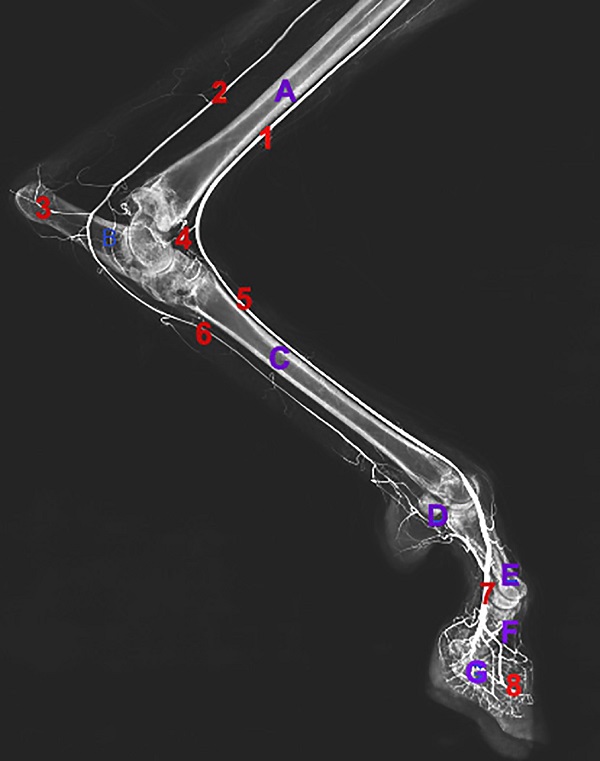
Fig. 1. Dorsal radiographic view of left hind limb in a goat (1: V. metatarsalis dorsali III; 2: V. digitalis dorsali communis; 3: Vv. digitalis dorsalis propria axialis). It was found that the remaining metatarsal veins united at the proximal third of the metatarsal into a single vein. This vein receives tributaries from the dorsum of dew claws, tarsal joint (Fig. 2. No. 4) which join the common dorsal digital vein (Fig. 2 No. 5) to continue as cranial tributary of V saphenous lateralis (Fig. 2. No. 1). On the planter aspect; the plantar venous plexus was composed of one to three large veins at the plantar digital area. It was observed that plantar proper digital vein (Fig. 2. No. 8) leads to the distal plantar arch just proximal to the fibrous pad on the fetlock joint, from, which the plantar common digital veins arise (Fig. 2. No. 6), after receiving tributaries from the proximal plantar arch continued as V. saphenous Medialis (Fig. 2. No. 2).
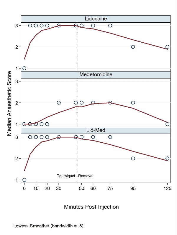
Fig. 2. Medial radiographic view of left hind limb in a goat (A: Tibia; B: Talus; C: Metatarsal bone; D: Proximal seasamoid bones; E: Proximal phalanx; F: Middle phalanx; G: Distal phalanx; 1: cranial tributary of V saphenous lateralis; 2: V. saphenous medialis; 3: Medial tarsal vein; 4: R. perforans tarsalis proximalis; 5: Dorsal common digital vein; 6: Vv. Digitalis plantaris communis; 7: V. digitalis dorsalis communis; 8: Vv. digitalis plantaris properis). Anesthetic evaluation The onset for desensitization was observed 12.0±6.0 seconds in LID and LID-MED groups. While it started in MED group at 4.0±2.0 minutes. In comparing with the baseline, the anaesthetic score was significantly higher in LID group (median = 3 at 5, 30 and 45 minutes and range = 2:3) before removal of elastic tourniquet (Fig. 3). After removal of elastic tourniquet, the anesthetic score decreased gradually (median = 2 at 5, 10 and 15 minutes, median = 1 at 30 minutes and range 1:3). In MED group the anesthetic score had a significant difference than the baseline. The median anesthetic score was 1 at 5, 15 and 20 minutes while it was 2 at 30 and 45 minutes (range=1:2) before removal of the elastic tourniquet. After removal of the elastic tourniquet the median anesthetic score was 2 at 5, 15 and 20 minutes and it was 1 at 30 minutes (range= 1:2). In LID-MED group the anesthetic score is significantly higher than the baseline before removal the elastic tourniquet (P = 0.038). The median anesthetic score was 3 before removal of elastic tourniquet (range = 2:3). After removal the elastic tourniquet, the median anesthetic score was 3 at 5, 15 and 20 minutes while it was 2 at 30 minutes (range = 2:3).
Fig. 3. Median anesthetic score at different times in LID, MED and LID-MED groups. At 5, 10 and 15 minutes after IVRA and before removal of the elastic tourniquet, MED group had a lower anesthetic score than LID and LID-MED groups (P = 0.034, 0.029 and 0.015 respectively). There were no significance differences between groups at the period started at 30 minutes after intravenous injection and lasts up to 30 minutes after removal of the elastic tourniquet. Regarding to cardiorespiratory variables, comparing with the baseline, in LID group; slight hyperthermia was observed at 30 and 45 minutes (P = 0.035 and 0.037 respectively) before removal of elastic tourniquet and at 20 and 30 minutes after removal of the elastic tourniquet (P = 0.01 and 0.005 respectively). Comparing data from the LID-MED group with the baseline revealed hypothermia at 30 and 45 minutes before removal of the elastic tourniquet. There were no significant differences in rectal temperature between all groups. In comparing with the baseline, marked bradycardia was observed in MED group at 15 minutes (P = 0.001). In comparing with the baseline and between groups, bradycardia was also observed in MED group at 20 minutes before removal of elastic tourniquet and lasts up to 30 minutes after removal of the elastic tourniquet. In LID-MED group, bradycardia than baseline and other groups was observed at 25 minutes post intravenous injection and lasts up to 30 minutes after removal of the elastic tourniquet. Increased respiratory rate in LID-MED group than the baseline and other groups was observed at 45 minutes before removal of elastic tourniquet and lasts up to 30 minutes after tourniquet removal (Table 2). Table 2. Time changes in cardiorespiratory valuables of goats subjected to intravenous regional anesthesia by lidocaine, medetomidine or lidocaine-medetomidine
Data are expressed as mean ± standard deviation in each group. In row, different small letter means significant P<0.05 while in each column, each capital letter means significant P<0.05. lidocaine (LID), medetomidine (MED), lidocaine-medetomidine (LID-MED). There was no sedation effect observed in LID group. The duration of sedation in MED group was 85.0±12.0 minutes while it was 91.0±10.0 minutes in LID-MED group without significant differences between these two groups. Sedation appeared in the form of recumbency, head directed backward and rest on abdomen, closed eye and salivation. Frequent urination was detected in MED and LID-MED groups. |
|||||||||||||||||||||||||||||||||||||||||||||||||||||||||||||||||||
|
|
|||||||||||||||||||||||||||||||||||||||||||||||||||||||||||||||||||
|
Discussion |
|||||||||||||||||||||||||||||||||||||||||||||||||||||||||||||||||||
|
The present study explored the clinical importance of the venous network of the pes region of goat's hind limb, which was agreed with previous studies on the terminal arch (Nickel et al., 1981 and konig and liebich, 2009). Moreover, blood drainage of the different veins in distal to proximal anatomical pattern (Dyce et al., 2010) was in the same line as proved in this article. The current study and other studies (Konig and liebich, 2009) were in agreement about the networking of the superficial and deep dorsal arches. The communication between planter and dorsal venous arches in the pes region enable obvious blood flow for the region that may aid in distribution of the injected local anesthetic. In the present study, the elastic tourniquet is left in its place for 45 minutes to avoid drug toxicity if the tourniquet was early removed and also to avoid local metabolic changes as hypoxia, hypercapnea, acidosis, hyperkalemia, and increased lactate concentration if the tourniquet remains for long time (Webb et al., 1999). Desensitization was rapidly appeared in LID and LID-MED groups. It may be due to the anesthetic effect of lidocaine. While slight desensitization was observed in MED group after longer time due to the analgesic effect of meditomidine (Tunio et al., 2003). Although adding dexmedetomidine to lidocaine for IVRA in human medicine may shorten the onset of sensory and motor block, improve postoperative analgesia and relief the pain of tourniquet (Memis et al., 2004), but in the present study the anesthetic effect in LID and LID-MED groups had no significant differences. Postoperative analgesia and pain induced by tourniquet were not evaluated in the present study, but the onset of desensitization was not significantly different between LID and LID-MED groups, which may be attributed to the small dose of medetomidine (20 µg/kg) used in the present study. The anesthetic effect remained up to 15 minutes after removal of the elastic tourniquet in both groups, which may be attributed to the slow absorption of anesthetic drug from the hind limb (Memis et al., 2004). Sedation was observed in MED and LID-MED groups as a consequence for the sedative effect of medetomidine (ὰ-2 agonists). Although slight, non-significant, anesthetic effect was noticed in the present study due to the effect of medetomidine IVRA, but further investigation is required to evaluate the analgesic effect, which was described in human medicine by Memis et al. (2004) and Esmaoglu et al. (2005). Slight significant hypothermia was detected in the present study in MED and LID-MED groups due to the systemic effect of medetomidine as previously mentioned by Carroll et al. (2005) and Tariq et al. (2011). Bradycardia was detected in MED and LID-MED groups after 15 minutes from intravenous injection and lasts up to 30 minutes post tourniquet removal. This bradycardia might be due to the effect of medetomidine (Hall et al., 2001; Carroll et al., 2005). In contrast with Carroll et al. (2005) and Tariq et al. (2011) the respiratory rate increased in the present study in MED and LID-MED groups. Shallow and rapid respiratory rate was noticed in the current study, which may be attributed to the lateral recumbency and ruminal stasis with gas production causing cranial pushing of the diaphragm. Monitoring the pressure of the elastic bandage is considered one of the limitations in the present study. |
|||||||||||||||||||||||||||||||||||||||||||||||||||||||||||||||||||
|
|
|||||||||||||||||||||||||||||||||||||||||||||||||||||||||||||||||||
|
Conclusion |
|||||||||||||||||||||||||||||||||||||||||||||||||||||||||||||||||||
|
Lidocaine-medetomidine combination used for intravenous regional anesthesia in goats gives satisfactory anesthesia. Anesthetic effect remains up to 15 minutes’ post tourniquet removal. Sedative effect is the additional effect of lidocaine-medetomidine combination than lidocaine alone. So according to anesthetic onset, anesthetic duration, anesthetic score and sedative effect LID-MED is considered the most reliable anesthetic combination used for surgical operation in pes region in goats. Further investigation is required for co-adjuvant intravenous regional anesthesia in veterinary practice in small ruminants. |
|||||||||||||||||||||||||||||||||||||||||||||||||||||||||||||||||||
|
|
|||||||||||||||||||||||||||||||||||||||||||||||||||||||||||||||||||
|
Conflict of Interests |
|||||||||||||||||||||||||||||||||||||||||||||||||||||||||||||||||||
|
Authors declared that no conflict of interest exist. |
|||||||||||||||||||||||||||||||||||||||||||||||||||||||||||||||||||
|
|
|||||||||||||||||||||||||||||||||||||||||||||||||||||||||||||||||||
|
References |
|||||||||||||||||||||||||||||||||||||||||||||||||||||||||||||||||||
|
|
|||||||||||||||||||||||||||||||||||||||||||||||||||||||||||||||||||
|
Acalovschi, I., Cristea, T., Margarit, S., Gavrus, R., 2001. Tramadol Added to Lidocaine for Intravenous Regional Anesthesia. Anesth. Analg. 92, 209–14. Babalolal, G.O., Oke, B. O., 1983. Intravenous regional analgesia for surgery of the limbs in goats. The veterinary quarterly 5, 186-189. Baggot D., 1982. Hoof lameness in dairy cattle. In practice 4, 133-141. Bigat, Z., Boztug, N., Hadimioglu, N., Cete, N., Coskunfirat, N., Ertok, E., 2006. Does Dexamethasone Improve the Quality of Intravenous Regional Anesthesia and Analgesia? A Randomized, Controlled Clinical Study. Anesth. Analg. 102, 605–9. Carroll, G.L., Hartsfield, S.M., Champney, T.H., Geller, S.C., Elizabeth, A., Martinez, E.A., Haley, E.L., 2005. Effect of medetomidine and its antagonism with atipamezole on stress-related hormones, metabolites, physiologic responses, sedation, and mechanical threshold in goats. Veterinary Anaesthesia and Analgesia 32, 147–157. Choyce A., Peng P., 2002. A systematic review of adjuncts for intravenous regional anesthesia for surgical procedures. Can. J. Anesth. 49, 32–45. Christodoulopoulos, G., 2009. Foot lameness in dairy goats. Res. Vet. Sci. 86, 281-284. Dyce, K.M., Sack, W.O., Wensing, C.J.G., 2010. Text book of veterinary anatomy (4th edn), Philaselphia, W. B. sounders, USA. El-Hawari, S.F., Sakata, H., Oyama, N., Tamura, J., Higuchi, C., Endo, Y., Miyoshi, K., Sano, T., Suzuki K., Yamashita K., 2018. Anesthetic and cardiorespiratory effects of single-bolus intravenous alfaxalone with or without intramuscular xylazine premedication in calves. Journal of Veterinary Medical Science 80, 361–367. Esmaoglu, A., Mizrak, A., Akin, A., Turk, Y., 2005. Addition of dexmedetomidine to lidocaine for intravenous regional anaesthesia. European Journal of Anaesthesiology 22, 447-451. Estill, C.T., 1977. Intravenous local analgesia of bovine lower leg. Veterinary medicine, small animal clinician 72, 1499-1502. Gentili, M., Bernard, J., Bonnet, F., 1999. Adding Clonidine to Lidocaine for Intravenous Regional Anesthesia Prevents Tourniquet Pain. Anesth. Analg. 88, 1327–30. Hall, L.W., Clarke, K.W., Trim, C.M., 2001. Veterinary Anaesthesia (10th edn), Harcourt, W. B. Saunders, London, UK. pp. 83–90. Holmes, C.M., 1963. Intravenous regional analgesia. A useful method of producing analgesia of the limbs. Lancet I, 245-247. Konig, H.E., liebich, H.G., 2009. Veterinary anatomy of domestic mammals. textbook and color atlas (4th edn), stutlgant, Schattauer Gmbh, Germany. pp. 474-482. Manohar, M., Kumar, R., Tyagi, R.P.S., 1971. Studies on intravenous retrograde regional anaesthesia of the forelimb in buffalo calves. Br. Vet. J. 127, 401-407. Memis, D., Turan, A., Karamanlıog, lu B., Pamukc¸u, Z., Kurt, I., 2004. Adding Dexmedetomidine to Lidocaine for Intravenous Regional Anesthesia. Anesth. Analg. 98, 835–40. Nickel, R., Schummer, A., Seiferle, E., 1981 The anatomy of domestic animals. The circulatory system, the skin and the cutaneous organs of domestic mammals. Vol.3, springer, Berlin, Germany. pp. 254-256. Prentice, D.E., Wyn-Jones, G., Jones, R.S., Jagger, D.W., 1974. Intravenous regional anaesthesia of the bovine foot. Veterinary Record 94, 293-295. Selda, S., Bakiye, U., Osman, N.A., Mustafa, O., Feray, G., Oner, S., 2006. The Analgesic Effect of Nitroglycerin Added to Lidocaine on Intravenous Regional Anesthesia. Anesthesia and Analgesia 102, 916-920. Sen, S., Ugur, B., Aydın, O. N., Ogurlu, M., Gezer, E., Savk, O., 2006. The analgesic effect of lornoxicam when added to lidocaine for intravenous regional anaesthesia. British Journal of Anaesthesia 97, 408–413. Tariq, M., Kalhoro, A.B., Kachiwal, A.B., Akhtar, A., Niamatullah, M., 2011. Sedative effects of medetomidine on pulse rate, respiratory rate and body temperature in cattle calves. Pakistan Journal of Science 63, 111-114. Tunio, A.N., Kalhoro, A.B., Kathio, I.H., 2003. Sedative and analgesic effects of detomidine hydrochloride in goats. Pakistan Veterinary Journal 23, 143-149. Weaver, A.D., 1972. Intravenous local anesthesia of the lower limb in cattle. Journal of American Veterinary Medical Association 160, 55-57. Weaver, A.D., Bogan, J.A., 1982. Intravenous regional anaesthesia of the bovine foot. Veterinary Record 110, 21. Webb, A.A., Cantwell, S.L., Duke, T., Adkins, E., 1999. Intravenous regional anesthesia (Bier block) in a dog. Can. Vet. J. 40, 419–442. Zabady M.K., Abu-Seida A.M., Ahmed K.A., 2004. Clinicopathological study on cutaneous squamous cell carcinoma and papilloma in sheep. Vet. Med. J., 589-600. |
|||||||||||||||||||||||||||||||||||||||||||||||||||||||||||||||||||